Real-Time Precision: Overcoming the Limitations of MRI in Neurosurgery
In stereotactic neurosurgery, procedures such as neuromodulation, biopsies, stereoelectroencephalography (sEEG), and lesioning demand sub-millimeter precision to achieve their intended therapeutic effect. However, current imaging methods—most commonly MRI (Magnetic Resonance Imaging)—pose significant limitations that impact surgical accuracy and outcomes. At Clee Medical, we have developed an intraoperative imaging system designed to overcome these challenges, enabling real-time neural targeting with micron-level precision.
The Problem: MRI and Its Inherent Limitations
- Resolution Constraints: standard MRI imaging, while widely used for surgical planning, typically provides a resolution of 1–2 mm. For procedures requiring extreme precision, such as brain biopsies or neuromodulation therapies, this resolution is insufficient—leaving surgeons to rely on estimations that introduce potential risk.
- Positional Inconsistencies: MRI images are taken pre-operatively with the patient in a specific position (usually lying down). However, during surgery, patient positioning changes—causing anatomical structures to shift. This discrepancy makes it harder to align pre-operative imaging with intraoperative reality.
- Brain Shift: once the skull is opened and brain surgery begins, the anatomy itself changes. This phenomenon, known as brain shift, occurs due to pressure changes, fluid loss, or tissue settling. Brain shift can displace critical structures by up to several millimeters—rendering pre-operative MRI plans increasingly inaccurate as the procedure progresses.
The result? Surgeons are faced with anatomical uncertainty and limited tools to adapt, which may compromise the accuracy of their interventions.
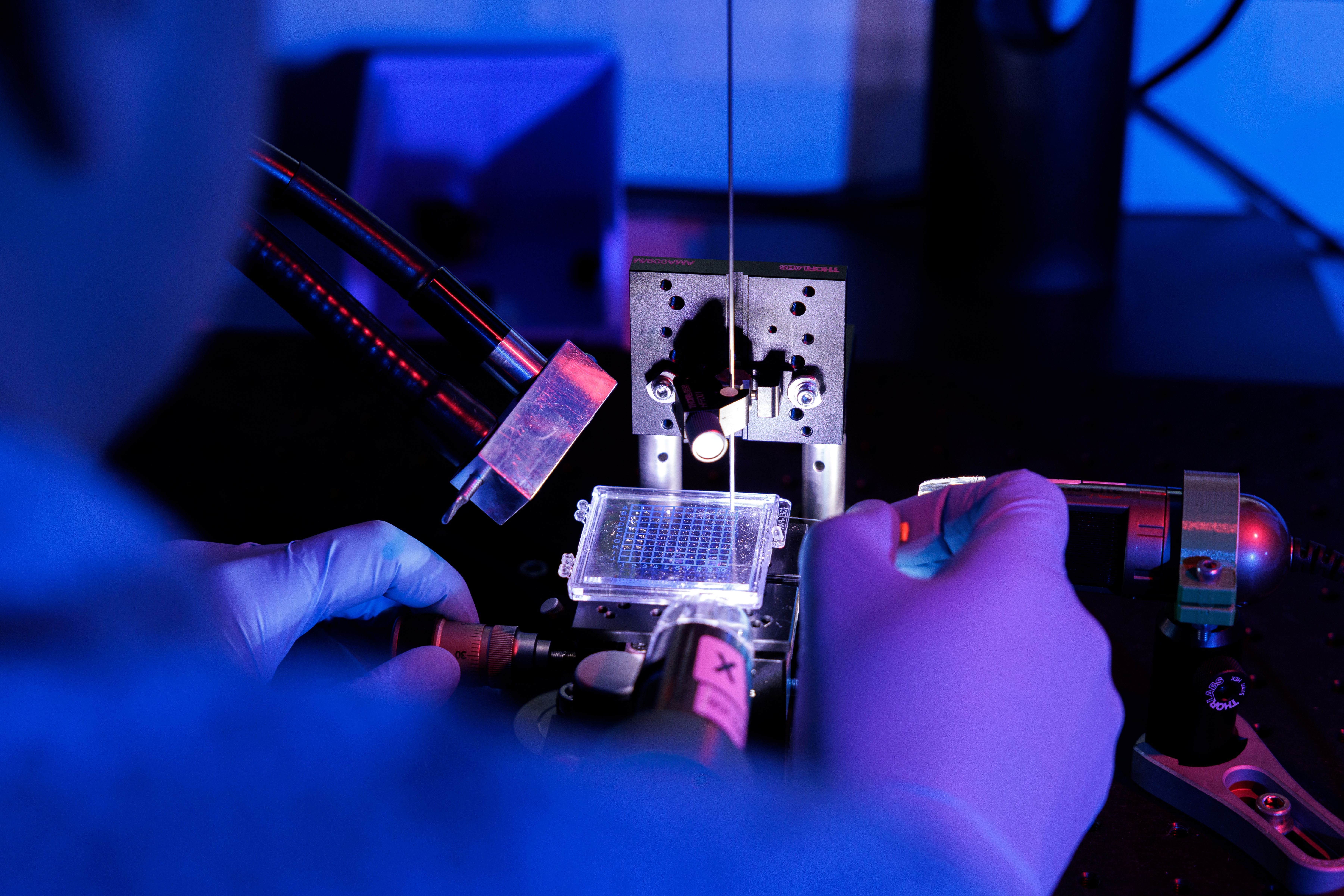
Clee Medical’s Intraoperative Imaging Solution
To address these challenges, we have developed an advanced intraoperative imaging system based on Optical Coherence Tomography (OCT) - a game-changing technology that provides live, high-resolution imaging of the brain during surgery. How it works:
- Real-Time Imaging: Unlike pre-operative MRI, OCT creates live images of brain anatomy during surgery, reflecting the true intraoperative conditions.
- Micron-Level Precision: our OCT solution creates unmatched clarity with micron-level resolution - enabling approximately 125,000 times more voxels per unit volume compared to traditional MRI.
- Radiation-Free and Contrast-Free: OCT imaging requires no radiation or contrast agents, ensuring safety and ease of use in the operating room.
- Focused Along the Surgical Trajectory: By concentrating imaging along the precise trajectory of the procedure, OCT enables surgeons to make confident, real-time targeting decisions.
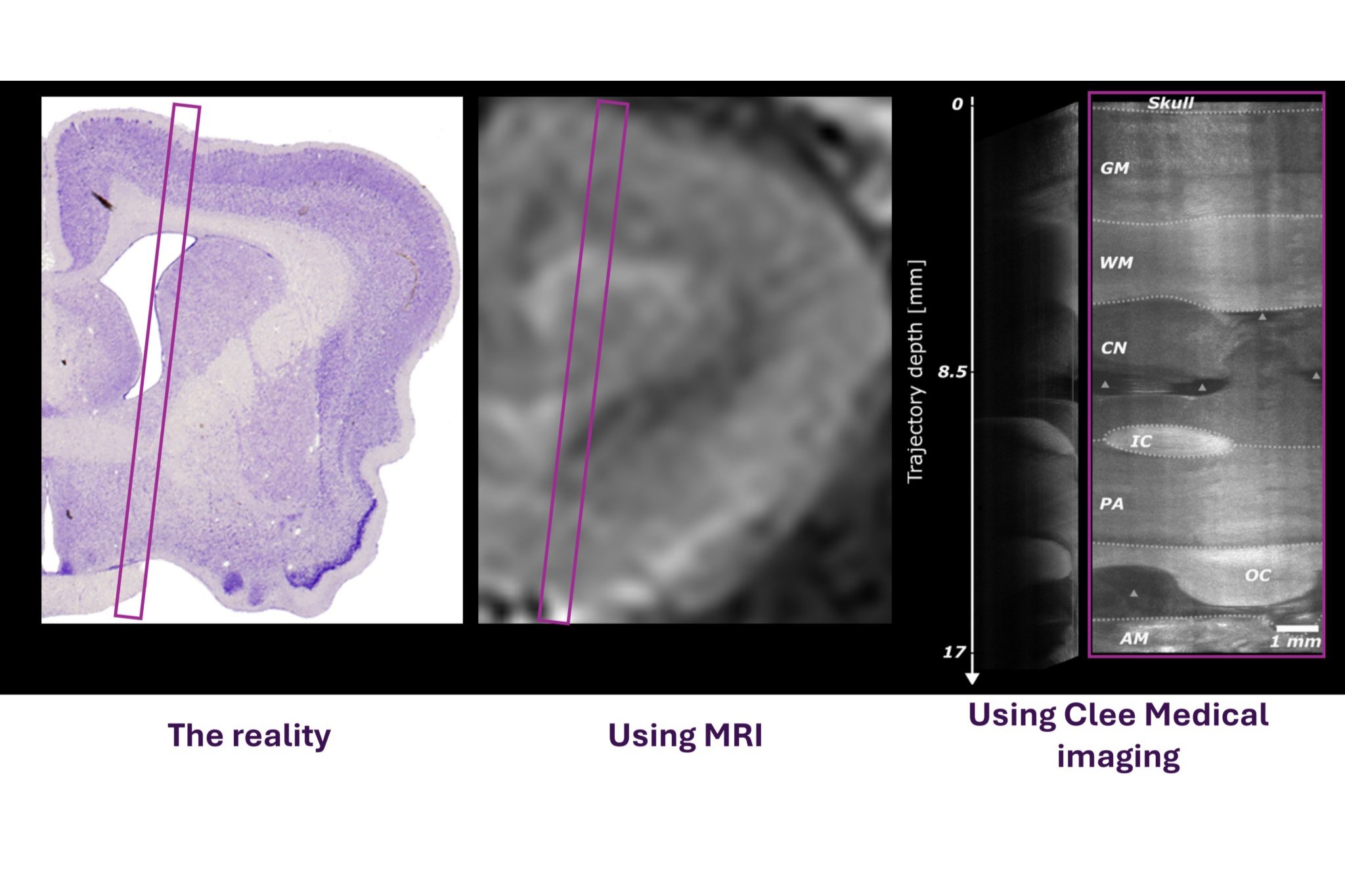
Live Animal Testing: Results that Speak for Themselves
In a live animal study conducted in 2024, we compared our intraoperative OCT imaging to standard pre-operative MRI and validated the findings using histology data (sourced from here as the gold standard atlas).
What we observed:
- Our OCT system enabled visualization of fine brain structures that could only be estimated using MRI.
- The micron-level clarity of OCT provided precise, real-time guidance that aligns with the actual brain anatomy during surgery—rather than relying on pre-operative estimations.
These findings demonstrate the transformative potential of our technology to support sub-millimeter neural targeting, even in the face of brain shift and anatomical variability.
The Impact: Precision, Confidence, and Therapeutic Effectiveness
With Clee Medical’s OCT-based imaging system, surgical teams gain the confidence they need to navigate the complexities of brain surgery with precision. By eliminating reliance on static MRI scans and addressing intraoperative changes in anatomy, our technology enables:
- Real-Time Decision Making: Surgeons can adapt to intraoperative challenges with live, accurate imaging.
- Sub-Millimeter Accuracy: Precision targeting ensures procedures are faster, safer, and more effective.
- Enhanced Therapeutic Outcomes: Patients receive the high level of care they deserve, with improved targeting accuracy leading to better clinical results.
Forging a New Standard in Neurosurgical Precision
The results of our testing underscore the promise of our intraoperative imaging system to revolutionize brain surgery. By providing surgeons with real-time, high-resolution visualization, we are closing the gap between pre-operative planning and intraoperative reality—offering unprecedented accuracy in neural targeting.
This is more than a step forward; it’s the foundation for a new era of precision-based brain treatments.